Using the figure above, identify the labeled part.
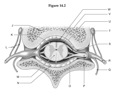
1. Label J: ______________________________
2. Label K: ______________________________
3. Label L: ______________________________
4. Label M: ______________________________
5. Label N: ______________________________
6. Label O: ______________________________
7. Label P: ______________________________
8. Label Q: ______________________________
9. Label R: ______________________________
10. Label S: ______________________________
11. Label T: ______________________________
12. Label U: ______________________________
13. Label V: ______________________________
14. Label W: ______________________________
1. Vertebral body
2. Pia mater
3. Rami communicantes
4. Spinal cord
5. Adipose tissue in epidural space
6. Denticulate ligament
7. Dorsal root ganglion
8. Dorsal ramus
9. Ventral ramus
10. Ventral root of spinal nerve
11. Autonomic (sympathetic) ganglion
12. Subarachnoid space
13. Arachnoid mater
14. Dura mater
You might also like to view...
The superior oblique eye muscles receive motor fibers from the __________ nerve.
Fill in the blank(s) with the appropriate word(s).
Aunt Jessie woke up one morning with excruciating pain in her chest. She had trouble breathing for several weeks. Following a visit to the doctor, she was told she had pleurisy. What is this condition and what did it affect?
What will be an ideal response?
What forms the matrix of blood?
What will be an ideal response?
The pre-embryonic period ends when
A. the blastocyst implants in the uterus. B. a morula is formed. C. the morula develops a trophoblast. D. the zygote undergoes cleavage.