Using the figure above, identify the labeled part.
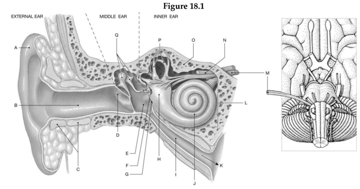
1. Label A: ______________________________
2. Label B: ______________________________
3. Label C: ______________________________
4. Label D: ______________________________
5. Label E: ______________________________
6. Label F: ______________________________
7. Label G: ______________________________
8. Label H: ______________________________
9. Label I: ______________________________
10. Label J: ______________________________
11. Label K: ______________________________
12. Label L: ______________________________
13. Label M: ______________________________
14. Label N: ______________________________
15. Label O: ______________________________
16. Label P: ______________________________
17. Label Q: ______________________________
1. Auricle
2. External acoustic meatus
3. Elastic cartilage
4. Tympanic membrane
5. Tympanic cavity
6. Oval window
7. Round window
8. Vestibule
9. Auditory tube
10. Cochlea
11. To nasopharynx
12. Bony labyrinth of inner ear
13. Vestibulocochlear nerve (N VII)
14. Facial nerve (N VII)
15. Petrous portion of temporal bone
16. Semicircular canals
17. Auditory ossicles
You might also like to view...
Where does hemopoiesis occur?
A. Epiphyseal line B. Endosteum C. Red bone marrow D. Yellow bone marrow E. Nutrient foramina
Which layer(s) of the wall of the GI tract contain a nerve plexus?
A. Muscularis only B. Adventitia only C. Submucosa and muscularis D. Mucosa and muscularis E. Mucosa and serosa
Functionally, a gomphosis is categorized as a
A. synovial joint. B. diarthrosis. C. synarthrosis. D. cartilagenous joint.
The ________ root carries sensory nerve fibers to the spinal cord.
Fill in the blank(s) with the appropriate word(s).