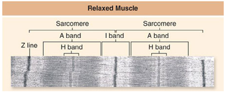
Shown below is an electron micrograph of part of a myofibril from a relaxed muscle fiber. Which of the labeled regions will NOT shorten when the muscle contracts?

A. A band
B. Sarcomere
C. H band
D. I band
Clarify Question
· What is the key concept addressed by the question?
· What type of thinking is required?
Gather Content
· What do you know about the structure of a myofibril? What other information is related to the question?
Choose Answer
· Given what you now know, what information is most likely to produce the correct answer?
Reflect on Process
· Did your problem-solving process lead you to the correct answer? If not, where did the process break down or lead you astray? How can you revise your approach to produce a more desirable result?
A. A band
Clarify Question
· What is the key concept addressed by the question?
o The question asks about the structure of a myofibril.
· What type of thinking is required?
o You are being asked to apply your knowledge about the structure of a myofibril to identify which will not change in shape during contraction.
Gather Content
· What do you know about the structure of a myofibril? What other information is related to the question?
o The A band is determined by the length of the thick filaments made up of myosin molecules in a sarcomere: this does not change length during a contraction. The Z lines form the borders of each sarcomere. The thin filaments are within the I bands and extend into the A bands interdigitated with thick filaments. The H band is the lighter-appearing central region of the A band containing only thick filaments. In a contracted muscle the Z lines have moved closer together, with the I bands and H bands becoming shorter. The A band does not change in size as it contains the thick filaments, which do not change in length.
Choose Answer
· Given what you now know, what information is most likely to produce the correct answer?
o The I band, H band and sarcomere become shorter during a muscle contraction as the actin and myosin filaments slide past each other. The A band does not change in length as it is defined by the myosin thick filament.
Reflect on Process
· Did your problem-solving process lead you to the correct answer? If not, where did the process break down or lead you astray? How can you revise your approach to produce a more desirable result?
o This question asked you to apply your knowledge about the structure of a myofibril to identify which will not change in shape during contraction. If you got the correct answer, great job! If you got an incorrect answer, where did the process break down? Did you think that the myosin molecules in the A chain shorten during a contraction? Did you think that actin and myosin sliding across each other would not affect the I band, H band, or sarcomere length?
You might also like to view...
These are the smallest functional units of matter that form all chemical substances and that cannot be further broken down by ordinary chemical or physical means.
A. protons B. neutrons C. electrons D. atoms E. molecules
To control microbial growth in a food, one can decrease water availability by the addition of ________.
A. agar B. gelatin C. sugar or salt D. formaldehyde
Which of the following types of membrane proteins are responsible for conveying external messages such as those sent by a hormone signal?
A) Recognition proteins
B) Receptor proteins
C) Connection proteins
D) Transport proteins
E) Enzymes
daughter cells are
What will be an ideal response?