Using the figure above, identify the labeled part.
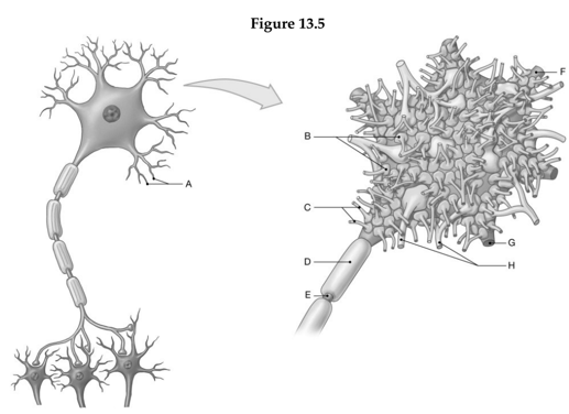
1. Label A: ______________________________
2. Label B: ______________________________
3. Label C: ______________________________
4. Label D: ______________________________
5. Label E: ______________________________
6. Label F: ______________________________
7. Label G: ______________________________
8. Label H: ______________________________
1. Dendrites
2. Terminal boutons
3. Terminal arborizations
4. Myelin sheath
5. Axon
6. Dendrite (cut)
7. Dendrite (cut)
8. Dendrite (cut)
You might also like to view...
What type of tissue comprises the valves of the heart?
a) Dense connective tissue b) Areolar connective tissue c) Hyaline cartilage d) Cardiac muscle tissue e) Adipose tissue
The __________________ ___________________ of the nephron acts as a countercurrent multiplier.
Fill in the blank(s) with the appropriate word(s).
The general name of the molecules given to an Rh negative individual to prevent the formation of Rh antibodies is ________.
Fill in the blank(s) with the appropriate word(s).
Where is cardiac muscle tissue located?
A) attached to bones B) heart C) tendons and ligaments D) ducts of certain glands