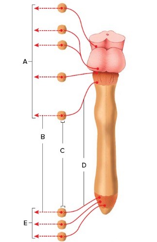
 The figure illustrates the parasympathetic division. What does "D" represent?
A. Cranial nerves
B. Pelvic splanchnic nerves
C. Postganglionic neurons
D. Preganglionic neurons
E. Terminal ganglia
Answer: D
You might also like to view...
The TMJ (temporomandibular joint) is composed of the convex upper condyle of the mandible, which fits into a concave surface of the temporal bone. The joint allows the mandible to swing uniaxially, producing the up-and-down motion of chewing. The TMJ is classified as a ________ joint.
Fill in the blank(s) with the appropriate word(s).
The output energy of all receptors is a type of ________ energy.
A. thermal B. nuclear C. mechanical D. chemical E. electrical
In the cerebral cortex, which cells process information on a local level?
A) Pyramidal B) Ependymal C) Stellate D) Purkinje
The neurotransmitter most likely produced when a person uses a drug that creates a sense of well-being is
A. glutamic acid. B. enkephalin. C. dopamine. D. substance Q.