Using the figure below, identify the labeled part.
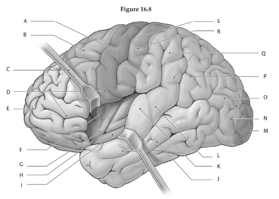
1) Label A: ______________________________
2) Label B: ______________________________
3) Label C: ______________________________
4) Label D: ______________________________
5) Label E: ______________________________
6) Label F: ______________________________
7) Label G: ______________________________
8) Label H: ______________________________
9) Label I: ______________________________
10) Label J: ______________________________
11) Label K: ______________________________
12) Label L: ______________________________
13) Label M: ______________________________
14) Label N: ______________________________
15) Label O: ______________________________
16) Label P: ______________________________
17) Label Q: ______________________________
18) Label R: ______________________________
19) Label S: ______________________________
1) Primary motor cortex (precentral gyrus
2) Somatic motor association area (premotor cortex
3) Retractor
4) Frontal lobe (retracted to show insula
5) Prefrontal cortex
6) Gustatory cortex
7) Insula
8) Lateral sulcus
9) Olfactory cortex
10) Temporal lobe (retracted to show olfactory cortex
11) Auditory cortex
12) Auditory association area
13) Visual cortex
14) Occipital lobe
15) Visual association area
16) Somatic sensory association area
17) Parietal lobe
18) Primary sensory cortex (postcentral gyrus
19) Central sulcus
You might also like to view...
The coccyx is typically composed of ________ fused vertebrae.
A) 1-2 B) 3-5 C) 6-7 D) 7-8 E) 9-11
During muscle contraction, myosin heads attach to actin and thereby form connections called ________.
Fill in the blank(s) with the appropriate word(s).
Which layer of the epidermis is found in only a few areas of the body?
A. Stratum corneum B. Stratum basale C. Stratum spinosum D. Stratum granulosum E. Stratum lucidum
which triangular muscle, located in the upper part of the chest, arises by three heads from ribs 3 to 5 converges on the shoulder?
a) trapezius b) deltoid c) rhomboid major d) pectoralis minor