Using the figure above, identify the labeled part.
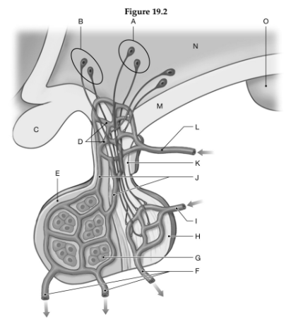
1. Label A: ______________________________
2. Label B: ______________________________
3. Label C: ______________________________
4. Label D: ______________________________
5. Label E: ______________________________
6. Label F: ______________________________
7. Label G: ______________________________
8. Label H: ______________________________
9. Label I: ______________________________
10. Label J: ______________________________
11. Label K: ______________________________
12. Label L: ______________________________
13. Label M: ______________________________
14. Label N: ______________________________
15. Label O: ______________________________
1. Paraventricular nuclei
2. Supraoptic nuclei
3. Optic chiasm
4. Capillary beds
5. Adenohypophysis of pituitary gland
6. Hypophyseal veins
7. Endocrine cells
8. Neurohypophysis of pituitary gland
9. Inferior hypophyseal artery
10. Portal veins
11. Infundibulum
12. Superior hypophyseal artery
13. Median eminence
14. Hypothalamus
15. Mamillary body
You might also like to view...
The ________ is the base, or floor, of the mouth.
A. tongue B. soft palate C. labia D. hard palate
While having a physical examination, a young male informed his doctor that at age 8 he had lobar pneumonia and pleurisy in his left lung. The physician decided to measure his VC. Describe the apparatus and method used for taking this measurement. Define the following terms used in the description of lung volumes: TV, IRV, ERV, RV, and VC.
What will be an ideal response?
The area of a myofibril where there are no actin filaments is the
A) H band. B) M line. C) I band. D) Z line. E) A band
Which of the following does NOT contribute secretions to seminal fluid?
A. Urinary bladder B. Seminal vesicles C. Bulbourethal glands D. Prostate gland