Using the figure below, identify the labeled part.
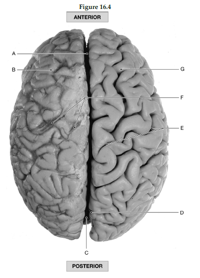
1) Label A: ______________________________
2) Label B: ______________________________
3) Label C: ______________________________
4) Label D: ______________________________
5) Label E: ______________________________
6) Label F: ______________________________
7) Label G: ______________________________
1) Longitudinal fissure
2) Left cerebral hemisphere
3) Cerebellum
4) Parieto-occipital sulcus
5) Central sulcus
6) Cerebral veins and arteries covered by arachnoid mater
7) Right cerebral hemisphere
You might also like to view...
Protrusion of an organ through a structure that normally contains it is referred to as a
a) hernia. b) goiter. c) strain. d) sprain. e) hydrocele.
Preganglionic neurons of the sympathetic division originate in the
A. dorsal horns of the craniosacral brain and spinal cord. B. ventral horns of the cervical and sacral spinal cord. C. dorsal horns of the thoracolumbar spinal segments. D. lateral horns of the thoracolumbar spinal segments. E. ventral horns of the cervical and thoracic spinal segments.
The __________ division tends to prepare the body for action.
A. sensory afferent B. motor afferent C. somatic motor D. parasympathetic E. sympathetic
A scientist genetically engineers a neuron whose voltage-gated Na+ channels lack the inactivation gate but which is otherwise normal. This neuron would be expected to
a. conduct action potentials bidirectionally. b. lack an absolute refractory period. c. lack a relative refractory period. d. all of these.