Using the figure below, identify the labeled part.
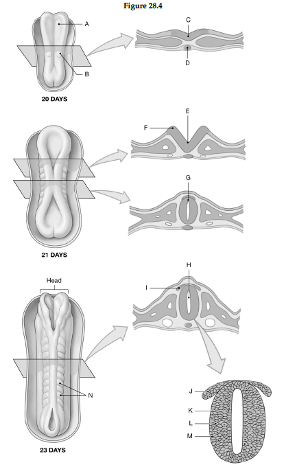
1) Label A: ______________________________
2) Label B: ______________________________
3) Label C: ______________________________
4) Label D: ______________________________
5) Label E: ______________________________
6) Label F: ______________________________
7) Label G: ______________________________
8) Label H: ______________________________
9) Label I: ______________________________
10) Label J: ______________________________
11) Label K: ______________________________
12) Label L: ______________________________
13) Label M: ______________________________
14) Label N: ______________________________
1) Neural plate
2) Somite
3) Neural plate
4) Notochord
5) Neural groove
6) Neural fold
7) Neural tube
8) Neurocoel
9) Neural crest
10) Neural crest
11) Ependymal layer
12) Mantle layer
13) Marginal layer
14) Somites
You might also like to view...
The cervical spinal cord
A) is smaller in diameter than other regions of the spinal cord. B) contains a smaller amount of gray matter than in other areas of the spinal cord. C) supplies the shoulder girdle and upper limb. D) All of the answers are correct. E) None of the answers are correct.
Answer the following statements true (T) or false (F)
1) Inflammation of the serous membranes of the abdominopelvic cavity is called peritonitis. 2) Inflammation caused by erosion due to gastric reflux is called a peptic ulcer. 3) Blockage of the lower part of the esophagus due to weak peristalsis and malfunction of the lower esophageal sphincter is called esophagitis. 4) Chief cells produce pepsinogen. 5) Parietal cells produce intrinsic factor.
Which are components of a hepatic portal triad? Choose all that apply.
A. Branch of hepatic artery B. Branch of hepatic portal vein C. Bile duct D. Branch of hepatic vein
Cardiac muscle cells ________
A) None of the listed responses is correct. B) lack striations C) have more than one nuclei per cell D) interconnect at intercalated discs