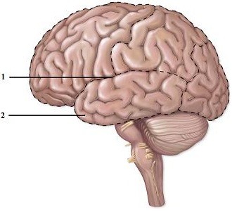
 This figure shows a left lateral view of the brain. What structure does number 1 indicate?
A. Lateral sulcus
B. Longitudinal fissure
C. Central sulcus
D. Lateral fissure
E. Central gyrus
Answer: A
You might also like to view...
Match the cell type to its tissue or function.
1. adipocyte 2. fibroblast 3. chondrocyte 4. osteocyte 5. osteoclast A. cartilage B. destroys bone matrix C. loose connective tissue D. fat E. maintains bone matrix
All of the following statements are true of inspiration except:
A. the rib cage is elevated. B. the diaphragm is relaxed. C. volume in the thoracic cavity has increased. D. intrapulmonary pressure has decreased.
The structure that connects the auricle to the tympanic membrane is called the ________
A) external acoustic meatus B) lobule C) vestibule D) helix
The auditory ossicle called the "anvil" is also known as the ________
A) malleus B) incus C) stapes D) bony labyrinth E) cochlea