Using the figure below, identify the labeled part.
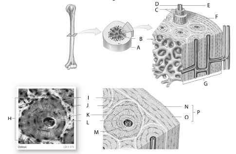
1 Label A: ______________________________
2 Label B: ______________________________
3 Label C: ______________________________
4 Label D: ______________________________
5 Label E: ______________________________
6 Label F: ______________________________
7 Label G: ______________________________
8 Label H: ______________________________
9 Label I: ______________________________
10 Label J: ______________________________
11 Label K: ______________________________
12 Label L: ______________________________
13 Label M: ______________________________
14 Label N: ______________________________
15 Label O: ______________________________
16 Label P: ______________________________
1 Compact bone
2 Spongy bone
3 Concentric lamellae
4 Capillary
5 Small vein (contained in central canal)
6 Periosteum
7 Compact bone
8 Osteon
9 Canaliculi
10 Osteocytes in lacunae
11 Matrix
12 Central canal
13 Blood vessels
14 Fibrous layer
15 Cellular layer
16 Periosteum
You might also like to view...
This figure shows a renal corpuscle. What structure does number 6 indicate?
A. Efferent arteriole B. Distal convoluted tubule C. Proximal convoluted tubule D. Afferent arteriole E. Nephron loop
Primary __________ occurs when an endocrine gland is secreting too little of its hormone because of an abnormality within that gland
Fill in the blank(s) with correct word
The portion of the kidney that is composed of cone-shaped renal pyramids is called the ________.
A. calyx B. pelvis C. columns D. cortex E. medulla
You are about to examine a blood smear from a patient who is suspected of being infected by a parasitic worm. What white blood cells would be present in elevated numbers due to such an infection?
A) Eosinophils B) Neutrophils C) Lymphocytes D) Basophils