Using the figure below, identify the labeled part.
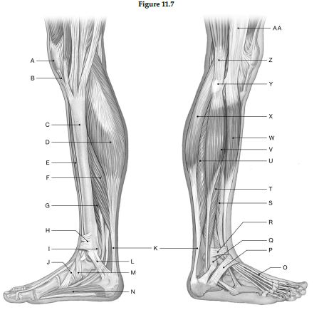
1) Label A: ______________________________
2) Label B: ______________________________
3) Label C: ______________________________
4) Label D: ______________________________
5) Label E: ______________________________
6) Label F: ______________________________
7) Label G: ______________________________
8) Label H: ______________________________
9) Label I: ______________________________
10) Label J: ______________________________
11) Label K: ______________________________
12) Label L: ______________________________
13) Label M: ______________________________
14) Label N: ______________________________
15) Label O: ______________________________
16) Label P: ______________________________
17) Label Q: ______________________________
18) Label R: ______________________________
19) Label S: ______________________________
20) Label T: ______________________________
21) Label U: ______________________________
22) Label V: ______________________________
23) Label W: ______________________________
24) Label X: ______________________________
25) Label Y: ______________________________
26) Label Z: ______________________________
27) Label AA: ______________________________
1) Patella
2) Patellar ligament
3) Medial surface of tibial shift
4) Medial head of gastrocnemius
5) Tibialis anterior
6) Soleus
7) Tibialis posterior
8) Superior extensor retinaculum
9) Medial malleolus
10) Tendon of tibialis anterior
11) Calcaneal tendon
12) Flexor retinaculum
13) Inferior extensor retinaculum
14) Abductor hallucis
15) Tendon of extensor hallucis longus
16) Inferior extensor retinaculum
17) Lateral malleolus
18) Superior extensor retinaculum
19) Extensor digitorum longus
20) Fibularis brevis
21) Soleus
22) Fibularis longus
23) Tibialis anterior
24) Lateral head of gastrocnemius
25) Head of fibula
26) Biceps femoris
27) Iliotibial tract
You might also like to view...
Which structure will sometimes disappear in adults?
A) Spleen B) Sinus C) Thymus D) Pancreas
Structure F is the
A) sphenoid bone. B) zygomatic process. C) temporal bone. D) zygomatic bone. E) middle concha.
The receptors that are stimulated by a large drop in the blood level are located in
a. the respiratory center of the brain b. carotid and aortic bodies c. tissue capillaries d. both b and c e. all of the above
During isovolumetric systole, pressure is highest in the __________.
a. left ventricle b. aorta c. pulmonary veins d. left atrium