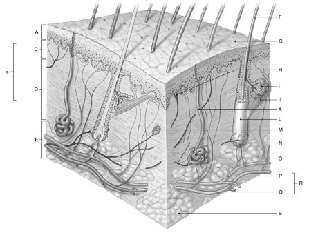
Using the figure below, identify the labeled part.

1 Label A: ______________________________
2 Label B: ______________________________
3 Label C: ______________________________
4 Label D: ______________________________
5 Label E: ______________________________
6 Label F: ______________________________
7 Label G: ______________________________
8 Label H: ______________________________
9 Label I: ______________________________
10 Label J: ______________________________
11 Label K: ______________________________
12 Label L: ______________________________
13 Label M: ______________________________
14 Label N: ______________________________
15 Label O: ______________________________
16 Label P: ______________________________
17 Label Q: ______________________________
18 Label R: ______________________________
19 Label S: ______________________________
1 Epidermis
2 Dermis
3 Papillary layer
4 Reticular layer
5 Subcutaneous layer (hypodermis
6 Hair shaft
7 Pore of sweat gland duct
8 Tactile corpuscle
9 Sebaceous gland
10 Arrector pili muscle
11 Sweat gland duct
12 Hair follicle
13 Lamellated corpuscle
14 Nerve fibers
15 Sweat gland
16 Artery
17 Vein
18 Cutaneous plexus
19 Fat
You might also like to view...
The radial fossa is seen on a coronal sectional image of the elbow lateral to the medial epicondyle and proximal to the trochlea of the distal humerus.
Answer the following statement true (T) or false (F)
List the eleven organ systems of the human body
What would be an ideal response?
Which of the following cell types is most numerous in the lung?
A. Squamous (type I) alveolar cells B. Great (type II) alveolar cells C. Ciliated columnar epithelial cells D. Goblet cells E. Alveolar macrophages
How does the breeding soundness evaluation of a stallion differ from that of a bull?
What will be an ideal response?