Using the figure below, identify the labeled part.
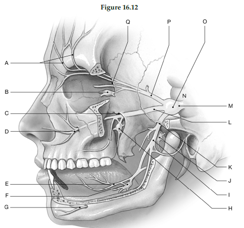
1) Label A: ______________________________
2) Label B: ______________________________
3) Label C: ______________________________
4) Label D: ______________________________
5) Label E: ______________________________
6) Label F: ______________________________
7) Label G: ______________________________
8) Label H: ______________________________
9) Label I: ______________________________
10) Label J: ______________________________
11) Label K: ______________________________
12) Label L: ______________________________
13) Label M: ______________________________
14) Label N: ______________________________
15) Label O: ______________________________
16) Label P: ______________________________
17) Label Q: ______________________________
1) Supraorbital nerves
2) Ciliary ganglion
3) Foramen rotundum
4) Infraorbital foramen
5) Lingual nerve
6) Submandibular ganglion
7) Mental nerve
8) Pterygopalatine ganglion
9) Mandibular branch
10) Otic ganglion
11) Foramen ovale
12) Maxillary branch
13) Trigeminal nerve (N V
14) Pons
15) Semilunar ganglion
16) Ophthalmic branch
17) Superior orbital fissure
You might also like to view...
The special functions of plasma and organelle membranes depend primarily on the specific composition of the phospholipids of those membranes.
Answer the following statement true (T) or false (F)
Which of the following is NOT a sign of pain?
a. vocal c. eating and drinking well b. restless d. chewing at incision
In response to the resultant rise in salt delivery to the distal tubule, the macula densa cells release __________ and adenosine
Fill in the blank(s) with correct word
Which movement involves raising the foot upward at the ankle joint?
a. abduction b. elevation c. dorsiflexion d. depression