Using the figure below, identify the labeled parts.
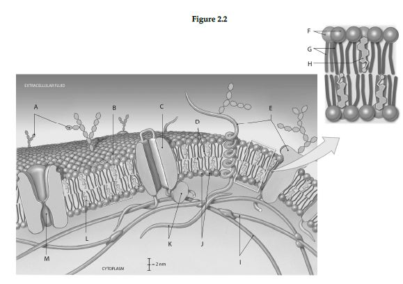
1 Label A: ______________________________
2 Label B: ______________________________
3 Label C: ______________________________
4 Label D: ______________________________
5 Label E: ______________________________
6 Label F: ______________________________
7 Label G: ______________________________
8 Label H: ______________________________
9 Label I: ______________________________
10 Label J: ______________________________
11 Label K: ______________________________
12 Label L: ______________________________
13 Label M: ______________________________
1 Glycolipids of glycocalyx
2 Phospholipid bilayer
3 Integral protein with channel
4 Hydrophobic tails
5 Integral glycoproteins
6 Hydrophilic heads
7 Hydrophobic tails
8 Cholesterol
9 Cytoskeleton (microfilaments
10 Hydrophilic heads
11 Peripheral proteins
12 Cholesterol
13 Gated chan
You might also like to view...
The ovarian cycle typically lasts about 28 days, with day 1 considered to be the first day after ovulation.
Answer the following statement true (T) or false (F)
A solution of a pH above 7 is called ________.
A. neutral B. isotonic C. acidic D. basic
The microscope of choice for a detailed three-dimensional study of the surface of a specimen is the
A. telescope. B. transmission electron microscope. C. light microscope. D. scanning electron microscope.
As a muscle gets stronger the diameter of its individual muscle cells becomes larger. Which of the following structures ensures that the membrane potential change is translated into the depth of the cell?
A. Z lines B. Transverse tubules C. Sarcoplasmic reticulum D. Pores in the plasma membrane E. H zone