Using the figure below, identify the labeled part.
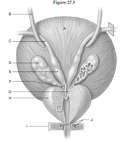
1) Label A: ______________________________
2) Label B: ______________________________
3) Label C: ______________________________
4) Label D: ______________________________
5) Label E: ______________________________
6) Label F: ______________________________
7) Label G: ______________________________
8) Label H: ______________________________
9) Label I: ______________________________
10) Label J: ______________________________
1) Urinary bladder
2) Ureter
3) Ductus deferens
4) Seminal gland
5) Ampulla of ductus deferens
6) Duct of seminal gland
7) Ejaculatory duct
8) Prostate gland
9) Urogenital diaphragm
10) Bulbo-urethral glands
You might also like to view...
There is a tremendous variety of signal transduction pathways with a wide range of functions. However, all these pathways have some things in common. Discuss some of these common features
What will be an ideal response?
It was often rumored that one of our deceased presidents was suffering from Addison's disease (inadequate synthesis of mineralocorticoids and glucocorticoids). What symptoms may have led to the diagnosis of this condition?
What will be an ideal response
Type II alveolar cells
A) allow rapid diffusion of gases through their thin membranes. B) allow rapid diffusion of gases through their thin membranes, secrete a chemical known as surfactant, and are phagocytic. C) secrete a chemical known as surfactant. D) are phagocytic. E) None of the statements are true.
The medullary cavity is
A. the site where osteoblasts are found. B. filled with fibrocartilage and elastin fibers. C. dead space in the bone. D. lined with endosteum. E. empty in adult bones.