Using the figure below, identify the labeled part.
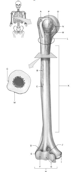
1) Label A: ______________________________
2) Label B: ______________________________
3) Label C: ______________________________
4) Label D: ______________________________
5) Label E: ______________________________
6) Label F: ______________________________
7) Label G: ______________________________
8) Label H: ______________________________
9) Label I: ______________________________
10) Label J: ______________________________
11) Label K: ______________________________
12) Label L: ______________________________
13) Label M: ______________________________
14) Label N: ______________________________
15) Label O: ______________________________
16) Label P: ______________________________
1) Greater tubercle
2) Deltoid tuberosity
3) Radial groove
4) Radial fossa
5) Lateral epicondyle
6) Capitulum
7) Trochlea
8) Condyle
9) Medial epicondyle
10) Coronoid fossa
11) Shaft (body
12) Surgical neck
13) Intertubercular sulcus
14) Anatomical neck
15) Head
16) Lesser tubercle
You might also like to view...
The ____________________ joint of the upper extremity has a fat pad.
Fill in the blank(s) with the appropriate word(s).
Our bodies need to coordinate and integrate all of their functions into one harmonious whole to maintain homeostasis.?
Indicate whether the statement is true or false
Stimulation of uterine muscle contractions during childbirth is triggered by ____
a. oxytocin b. prolactin c. estrogen d. aldosterone
Nodes of Ranvier are
A) interruptions in the myelin sheath along the course of a myelinated axon. B) collections of immune cells in the CNS. C) satellite cells that support neuron cell bodies in ganglia. D) gaps between choroids plexuses where cerebral spinal fluid emerges